filmov
tv
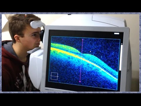
Normal macular ANAtomy on OCT
0:00:27
High risk proliferative diabetic retinopathy #eyedisease #retina #ophthalmology
0:09:02
Course in clinical applications of OCT for macular diseases - Neovascular ARMD
0:00:11
Choroidal Rupture #choroid #fundusexamination#ophthalmology #optometrists #diagnosis
0:00:14
Normal fundus | Normal Retina | Smartphone Fundus Videography | Fundus Photography | Short Video 86
0:00:15
Clear Image of Retina (Funduscopy), Aankh ka parda, Retinal layers , Macula, Optics disc
0:00:07
Normal Fundus Photo #shorts #retina #eyes #fundusphoto #viralshorts #macula@Eyecare_tips
0:00:18
Retinal Arteriolar Macroaneurysm
0:00:14
Drusen + hypertensive retinopathy #retina #oftalmo #ophthalmology #oftalmologia #oftalmología #oph
0:00:20
Stargardt disease
0:00:18
Branch retinal artery occlusion #retina #oftalmo #ophthalmology #oftalmologia #oftalmología #ophta
0:00:16
Optical coherence tomography #ophthalmology #optometrists #retinopathy
0:00:15
Flame shaped Hemorrhages | proliferative diabetic retinopathy PDR | Short Video 180
0:18:06
Age- related macular degeneration (AMD) | Pathogenesis of dry AMD and Wet AMD, drusens ,CNVM..
0:00:12
Branch Retinal Vein Occlusion (BRVO) #retina #ophthalmology #americanacademyofophthalmology
0:00:15
Diabetic retinopathy | cotton wool spots | Fundus | Short Video 172 #optometryacademy #akleshkumar
0:03:46
What is OCT Scanning? (Optical Coherence Tomography)
0:00:18
Disc oedema | Smartphone Fundus Videography | Fundus Photography | Short Video 33
0:00:53
Basic Eye Anatomy by Vicki Chan MD
0:00:25
Optic disc pit maculopathy
0:00:14
Normal fundus | Normal Retina | Smartphone Fundus Videography | Fundus Photography | Short Video 148
0:00:16
VALSALVA RETINOPATHY #optometrists #ophthalmology #mbbs #retinal
0:00:14
Normal fundus | Normal Retina | Smartphone Fundus Videography | Fundus Photography | Short Video 267
0:00:19
Anterior segment of eyeball includes structure laying infront of the... Optometry MCQ - Optometry fa
0:00:40
Wide field BLFI images and OCT in a case of polypoidal choroidal vasculopathy (PCV)
Назад
Вперёд
 0:00:27
 0:09:02
 0:00:11
 0:00:14
 0:00:15
 0:00:07
 0:00:18
 0:00:14
 0:00:20
 0:00:18
 0:00:16
 0:00:15
 0:18:06
 0:00:12
 0:00:15
 0:03:46
 0:00:18
 0:00:53
 0:00:25
 0:00:14
 0:00:16
 0:00:14
 0:00:19
 0:00:40